Shear wave research systems offer detailed insights into tissue elasticity and stiffness.
Shear waves are elastic waves that pass through tissues. The systems can create detailed elastography images that help in assessing the medical properties of tissues.
Let’s learn more about them below.
Components of a shear wave research system
It comprises several key components, including:
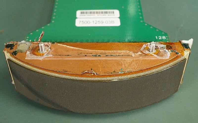
● Ultrasound transducer

This device generates and receives ultrasound waves. They make the shear waves and record their propagation through tissues.
The transducer delivers electrical signals into mechanical vibrations that travel through the body.
When the waves meet different tissues, they are reflected to the transducer. It later transforms them back into electrical signals for analysis.
● Acoustic radiation force (ARF) excitation

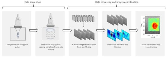
This component of the Shear wave research system generates the shear waves by applying a focused ultrasound beam to the tissue, causing localized displacement.
The ARF excitation creates the shear waves that spread through the tissue, allowing for the measurement of tissue stiffness.
This process uses high-intensity focused ultrasound to push on the tissue, making a shear wave that moves perpendicular to the direction of the ultrasound beam.
● Signal processing unit

This unit processes the delivered ultrasound signals to take measurements of the shear wave speed and regulate elastography images.
The signal processing unit uses advanced algorithms to check the time it takes for the shear wave to travel through the tissue.
The system can regulate the stiffness of the tissue with the measurement of the wave speed. It is then displayed as an elastography image.
● Display and analysis software

Healthcare providers use advanced software to check the elastography images generated by Shear wave research systems and examine the tissue stiffness. The software provides a user-friendly link between clinicians and researchers to decode the data.
It takes measurements and compares the tissue stiffness with a tool to store and share the images and data.
By having these components, this shear wave becomes the best research ultrasound system.
Applications of shear wave research systems
This system has various applications, including:
● Medical diagnostics
These systems are used to check:
- liver fibrosis
- breast lesions
- thyroid nodules by measuring tissue stiffness
● Musculoskeletal imaging
They help in
- analyzing muscle and tendon elasticity
- diagnosis of conditions like tendinopathy and muscle injury
Medical professionals use Shear wave research systems to gain knowledge about the mechanical properties of muscles and tendons.
This helps in the diagnose and monitor sports injuries and other musculoskeletal conditions.
● Cardiovascular research
Professionals use shear wave elastography as a way to measure the stiffness of the arteries. Doctors use this test to find check for diseases like atherosclerosis.
● Oncology
Shear wave research systems help to detect and characterize tumors by making differences between benign and malignant tissues based on their stiffness.
Tumors have different mechanical properties than the surrounding healthy tissues, and shear waves can help identify the differences.
These systems help in the diagnosis and treatment planning for cancer patients.
Why is it the best?
Shear wave research systems are considered the best due to several reasons:
● Non invasive and safe
They use a safe method to detect the tissue properties without the need for biopsy and other invasive procedures. It reduces the risk of complexities and discomfort for patients.
The non-invasive nature of shear wave elastography is an attractive option for routine screening and monitoring various conditions.
● High accuracy and reliability
The systems offer proper measurements of tissue stiffness, helping for proper diagnosis and monitoring.
The advanced signal processing algorithms and high quality transducers used in these Shear wave research systems make sure that the measurements are reliable and reproducible.
This accuracy is necessary to make informed decisions and track the progression of diseases.
● Versatility
They are used in various medical fields like hepatology, oncology etc. It makes them highly versatile.
The ability to determine tissue stiffness in different organs and tissues makes shear wave elastography a demanding tool for various applications.
The versatility also means that you can use the same system for various purposes, increasing its benefits and cost effectiveness.
● Real time imaging
The ability to deliver real time elastography images allows for immediate determination and making decisions during clinical examination.
Clinicians become able to see the results of the elastography scan instantly, which is useful in managing procedures like biopsy or in determining the instant effects of treatments.
Conclusion
Shear wave research systems represent a significant advancement in medical imaging technology.
The ability to provide detailed analysis of tissue stiffness makes them vital in many medical applications.
The combination of advanced technology, user friendly software, and real time imaging abilities make it the best research ultrasound system. These qualities also ensure that the research systems play an important role in the medical diagnostics and research.
Visit our home-page for more information


